Archives 2024

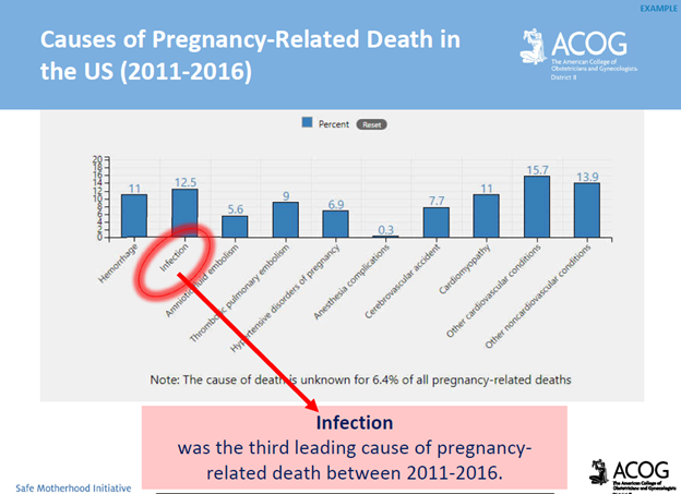
THE LINK BETWEEN UTERINE ACTIVITY IN LABOR, FETAL (IN)TOLERANCE, AND LIABILITY

In this blog, I’ll review basic uteroplacental physiology in labor, as well as the components of a complete uterine activity assessment.  I’ll highlight some litigation trends related to uterine activity in labor, and close with risk management approaches. 

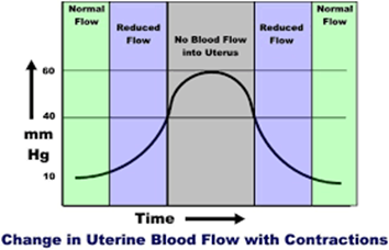
Physiology Basics: The uterus is a muscle that contracts in labor. Blood supply to the uterus, placenta, and fetus is dependent on maternal blood pressure, as well as uterine activity (e.g. contractions). With every labor contraction, the blood supply to the uterus, placenta, and fetus is reduced by 60%.

  • 75% of fetal hypoxia cases in the intrapartum period occur slowly, and progressively from the impact of uterine contractions over a period of time, rather than from an acute event.  
  • Labor contractions can reduce the fetal partial pressure of oxygen (PaO2) by 25%.  The healthy fetus is able to tolerate this, however, a high-risk fetus (e.g. fetus sustaining recurrent significant deceleration’s, fetal growth restriction, maternal history of chronic hypertension), may not be able to tolerate this reduction.
  • The relaxation time between contractions results in the ability for restoration of maternal-fetal gas exchange, and allows the fetus to return the PaO2 to normal levels, and clear carbon dioxide (CO2), and lactate.
    • Fetal lactate is a by-product of fetal anaerobic metabolism related to periods of hypoxia (low oxygen levels in the fetus’ body tissues.)

Fetal (In)Tolerance to Labor Contractions: Excessive uterine activity can have an adverse effect on fetal oxygenation, and the acid-base status of the fetus.  Excessive uterine activity can result in decreased fetal cerebral oxygen saturation, as well as fetal acidemia (fetal blood with high levels of acid, or a low pH).  Excessive uterine activity can result in birth injuries, specifically, hypoxic ischemic encephalopathy, subsequent cerebral palsy, and possible death.

Excessive uterine activity can have an adverse effect on uterine re-perfusion as well.  In the presence of anaerobic metabolism, elevated uterine lactate levels can develop.  This can result in inadequate labor contractions, ultimately leading to Pitocin augmentation, cesarean delivery, postpartum hemorrhage, and associated sequelae.  

The number one cause of fetal intolerance to labor contractions is inadequate re-perfusion due to inadequate relaxation time between contractions.

  • Uterine activity affects the fetal base deficit (BD)
    • There is a minimal effect from labor onset to 4cm dilation
    • From 4cm dilation to complete dilation, the mean BD increases by approximately 1mmol/L every 3 hours due to the increased frequency, and intensity of contractions
    • In second stage labor, the BD will increase by 1mmol/L every 1 hour due to the increased strength, and frequency of contractions, as well as maternal pushing efforts
    • The effects on BD are more significant with an interruption of the maternal fetal oxygen pathway (e.g. presence of variable, late, and/or prolonged decelerations), as well as with excessive uterine activity
  • Umbilical artery pH is reduced, while lactate levels are increased with increasing uterine activity, intrauterine pressure, and resting tones

Complete Assessment of Uterine Activity: The assessment of the fetus should not be performed in the absence of a complete assessment of uterine activity.  Components of uterine activity assessment, and documentation include – 

  • uterine contraction frequency: The number of contractions in a 10-minute period
    • normal frequency ranges from 2 – 5 contractions per 10 minutes
    • frequency alone is only a partial assessment of uterine activity
    • identified as abnormal with:
      • > 5 contractions in 10 minutes averaged over 30 minutes = uterine tachysystole
        • tachysystole includes spontaneous, and induced contractions
        • requires treatment even if the fetal heart rate (FHR) tracing is normal
Normal Uterine Activity
Tachysystole
  • duration of uterine contractions: The time from the onset of a contraction, to the offset of a contraction measured from baseline resting tone
    • ranges from 45 – 80 seconds
  • strength or intensity of uterine contractions: The peak of a contraction minus the resting tone
    •  25 – 50 mmHg (millimeters of mercury) in the first stage of labor
    •  may rise to >80 mmHg in the second stage of labor
    •  contractions palpated as “mild” would peak at < 50 mmHg if measured internally. Contractions palpated as “moderate” or greater would peak at 50 mmHg or greater if measured internally 
  • resting tone of uterus: The intrauterine pressure when the uterus is not contracting
    • the average resting tone during labor is 10 mmHg.  If assessment if performed by palpation (touching), assessment should palpate as “soft”: easily indented
    • increased resting tone = hypertonus
      • resting tone > 20-25mmHg, or  uterus does not palpate soft between contractions
Hypertonus
  • relaxation time between uterine contractions: The time-frame from the end of one contraction to the beginning of the next
    • this is usually 60 seconds or more in the first stage of labor, and 45 seconds or more in the second stage
    • frequently confused with resting tone
  • Montevideo units (MVUs): Assessed and documented if an intrauterine pressure catheter (IUPC) is in place.  The average intensity of contractions in mmHg multiplied by the number of contractions in 10 minutes
    • MVUs range from 100 – 250 MVUs in the first stage of labor.  MVUs may rise to 300 – 400 MVUs in the second stage of labor
    •  “adequate” labor contractions are defined as > 200 MVUs 

What is Excessive Uterine Activity? All definitions for excessive uterine activity apply to both spontaneous, and induced, or augmented labor. The evaluation of the presence, or absence of uterine tachysystole, in addition to evaluation of contraction duration, uterine resting tone, and relaxation time between contractions.

  1. Tachysystole: contraction frequency >5 in 10 minutes, averaged over 30 minutes.
  2. Hypertonus: uterine resting tone exceeding 20 -25 mmHg with an IUPC, or a uterus that doe not return to “soft” by palpation during relaxation between contractions.
  3. Inadequate relaxation time: less than 60 seconds of uterine relaxation between contractions during the first stage of labor; less than 45 seconds of uterine relaxation between contractions in the second stage of labor.
  4. Excessive contraction duration (i.e. prolonged contractions, tetanic contractions, uterine tetany.) A series of single contractions lasting 2 minutes or more.
Excessive Uterine Activity & Tachysystole

Management of Excessive Uterine Activity: Management of excessive uterine activity should not be based on the presence or absence of FHR changes. 

The goal of management is to identify, and promote normal uterine activity, and correct the underlying cause of any type of excessive uterine activity.

  • maternal position change to side-lying
  • administration of an intravenous fluid bolus
  • removal of cervical ripening agents, or decrease or discontinue the use of oxytocin
  • use of a tocolytic (i.e., terbutaline) if the above interventions were ineffective, or the excessive uterine activity occurs in the presence of FHR changes indicative of interrupted fetal oxygenation

EXCESSIVE UTERINE ACTIVITY SHOULD TRIGGER INTERVENTIONS, REGARDLESS OF THE FHR STATUS

Uterine Tachysystole, and Excessive Uterine Activity:  A Common Area of Liability

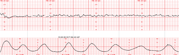
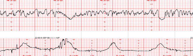
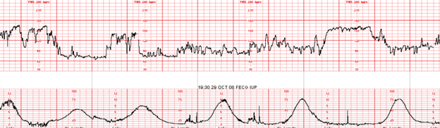
Case Study (10/07/2020)

A 28-year-old gravida 2, para 1 at 37 weeks gestation presented for a scheduled Pitocin induction of labor for preeclampsia without severe features.

Admission assessments were unremarkable, with a baseline favorable cervical examination of 3/80/-1. The labor course was complicated by a recurrent Category II FHR tracing in the presence of excessive uterine activity. The patient progressed to complete dilation, attempted to push with contractions, resulting in an onset of fetal bradycardia.  An emergency cesarean delivery occurred resulting in delivery of a viable male weighing 7#2oz with Apgar scores of 1/3/3/3/5. A venous cord gas revealed metabolic acidemia: pH 6.94, BD 14.6.  An arterial cord gas was unable to be obtained.

The newborn was diagnosed with hypoxic ischemic encephalopathy (HIE), and received therapeutic hypothermia. The newborn had an onset of seizures at seven hours of life. A brain MRI was performed on day of life five revealing a watershed pattern of injury.  The newborn was discharged home with the parents on day of life 16. The infant suffers from spastic cerebral palsy with profound neurodevelopmental delays.

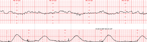
EFM Tracing with Annotated Segment of Ob Provider Note:

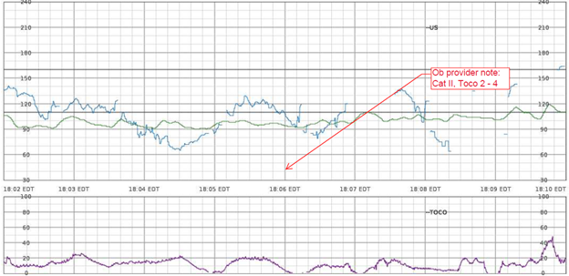
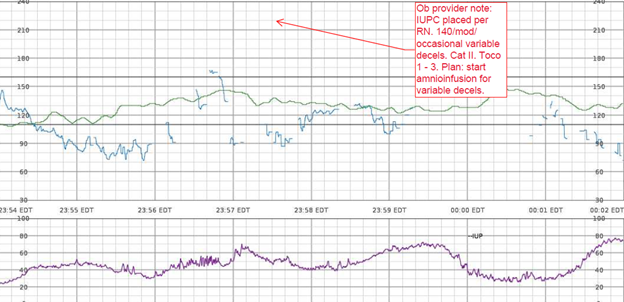
Nurses Notes: RN notes indicate a “normal” uterine contraction pattern, normal duration of contractions, and a “soft” resting tone throughout labor.

Q: What assessments are missing specific to uterine activity?

A: contraction duration, resting tone in mmHg, relaxation time between contractions, MVUs in the presence of an IUPC, uterine, and fetal responses to interventions

Litigation Case Theme: Failure of the perinatal team to treat a Category II FHR tracing in the presence of uterine tachysystole, and excessive uterine activity resulting in HIE, and subsequent permanent neurological injuries. 

Plaintiff Allegations:

Failure to appropriately identify and treat uterine tachysystole, and excessive uterine activity in the presence of a Category II FHR tracing in a timely manner

Failure to discontinue oxytocin in the presence of excessive uterine activity, and a Category II FHR tracing

Inappropriate oxytocin management

Failure to initiate intrauterine resuscitation

Failure to follow oxytocin orders, and policy

Failure to activate the chain of command when there was clinical disagreement between the nurse, and responsible physician

Defenses:

The fetal injury likely occurred during the antenatal period; therefore, actions during labor and delivery had no impact on the outcome

The standard of care was adhered to

Documentation reflected prompt, and appropriate actions by the perinatal team

Electronic fetal monitoring cannot be used as a diagnostic tool; therefore, birth injury cannot be attributed solely to FHR interpretation

Standard of Care Takeaways (Risk Management Approaches): the evaluation of uterine activity must occur, and is equally as relevant as the assessment of the fetal heart rate. 

  • Hospital systems should have clear definitions of tachysystole, and excessive uterine activity as clinical management, and policies/procedures should guide expected interventions when tachysystole or excessive uterine activity is identified.
  • Ensure organizational agreement on guidelines specific to uterine activity, not just management of tachysystole
  • All members of the perinatal team (physicians, residents, midwives, nurse practitioners, nurses) should be aware of the clinical criteria established for uterine tachysystole, and excessive uterine activity.
  • Most episodes of uterine tachysystole, and excessive uterine activity, occur as a result of the administration of oxytocin (60%). 
  • Treat uterine tachysystole, and excessive uterine activity by decreasing or discontinuing oxytocin, even in the presence of a Category I FHR tracing.
  • Avoid prolonged periods of uterine tachysystole, and excessive uterine activity that leads to progressive deterioration of the fetal status, and subsequent indeterminate (Category II) or abnormal (Category III) FHR patterns.
  • Treatment for uterine tachysystole, and excessive uterine activity should not be delayed until there is evidence of an indeterminate (Category II) or abnormal (Category III) fetal status.
  • All members of the perinatal team should be consistent with their assessments, interventions, and communications with colleagues.
  •  Nurses should advocate for patient safety if they feel pressured to increase oxytocin rates during uterine tachysystole and/or indeterminate (Category II) or abnormal (Category III) FHR patterns.
  • Remain current with your professional organization, and organizational literature
  • Consistently communicate, and document utilizing the standardized nomenclature including all characteristics of uterine activity assessment (frequency, intensity, duration, resting tone, relaxation time).
  • Ensure all members of the perinatal team are aware of the components of a complete assessment of uterine activity in labor.
  • Recognize that contraction frequency alone is a partial assessment of uterine activity.  Other components are equally important: duration, intensity, resting tone, relaxation time. Attain, and maintain adequate uterine activity.
  • Be able to identify signs of interruption in the maternal fetal oxygen pathway.

Conclusion: The assessment of uterine activity during labor is crucial, and is considered a patient safety issue.  Applying known parameters to the assessment of uterine activity influences management decisions, forms the basis of safe use of labor stimulants, and provides a means of defining excessive uterine activity among the multidisciplinary perinatal team.

The goal of this blog post is to improve birth outcomes, reduce liability risk to perinatal care providers, educate perinatal team members to avoid the risk of recurrence of errors, while supporting the reduction of obstetrical related medical malpractice lawsuits.

References:

AAP, ACOG (2017). Guidelines for Perinatal Care

ACOG (2009). Induction of Labor

ACOG (2010). Intrapartum Fetal Heart Rate Tracings

AWHONN (2014). Perinatal Nursing

AWHONN (2021). Perinatal Nursing

AWHONN (2022). Intermediate Fetal Monitoring Course

Miller et al., (2022). Mosby’s Pocket Guide to Fetal Monitoring

Rimsza et al., (2024). Association between Elevated Intrauterine Resting Tone during Labor and Neonatal Morbidity. American Journal of Perinatology

Ross et al., (2002). Use of umbilical artery base excess: algorithm for the timing of hypoxic injury. American Journal of Obstetrics & Gynecology

Turner et al., (2020). The physiology of intrapartum fetal compromise at term. American Journal of Obstetrics and Gynecology

COMMENT AND SHARE: What is your experience with a lawsuit involving uterine tachysystole, or excessive uterine activity? What was the case theme? Did the case go to trial? What was the case verdict?

DATA ABOUT DATA: What You Should Know

You can learn so much from the metadata in the electronic health record (EHR).  Metadata is data, about data.

Example: data entered, patterns of missing data, when data was entered, who entered it, who viewed it, how long it was viewed for, and whether it was modified.

Metadata can identify incidences of errors, as well as patterns of patient care delivery (i.e., recurrent late entries)

Metadata is discoverable per the Federal Rules of Civil Procedures. This means that attorneys can acquire access to EHR information, including the metadata, through the discovery process. Metadata is typically  obtained by a computer-generated record of audit trails showing user access and actions.  

Providers Can Minimize Risk by Effectively Documenting: During the litigation process, metadata can play an integral role in determining the credibility of evidence, including healthcare provider’s testimony, and documentation.

  • Avoid documentation gaps
  • Don’t copy and paste text from one patient’s EHR to another
  • Use templates and checklists cautiously
  • Do not share your password
  • Make any changes to the record as soon as possible, per organizational policy
  • Know that what you view is recorded
  • Document referrals and notifications of other nurses about changes in a patient’s condition

Metadata analysis can support — or not support — a lawsuit. Frequent errors, and errors of omission can negatively impact a healthcare providers credibility in court.  Contrary, metadata that demonstrates  complete, and accurate documentation can help exonerate healthcare providers by bolstering their credibility, and providing evidence that adherence to organizational policies, and procedures, as well as  standards of practice were followed.

RESOURCES

  • AHIMA. E-discover litigation and regulatory investigation response planning: Crucial components of your organization’s information and data governance processes. n.d. https://bok.ahima.org/doc?oid=107115#.ZByfmBXMJhE
  • Barrett M, DeAngelo TR, DeAngelo JG. E-discovery: Metadata analysis in medical malpractice litigation. The Legal Intelligencer. 2020. Commentary. https://www.law.com/thelegalintelligencer/2020/04/09/e-discovery-metadata-analysis-in-medical-malpractice-litigation/?slreturn=20230223103325
  • Conn J. Making IT legal-size; As electronic health-record systems become more complex, so do the issues involving the legal status of those records. Modern Healthcare. 2008;38(20),
  • Gardner E. The weight of the I.T. evidence; why EHRs won’t reduce your malpractice premiums. Health Data Management. 2013;21(10).
  • Hansen MD, Pratt TJ. Follow the audit trial: The impact of metadata in litigation. Defense Counsel J. 2017;84(3).
  • NSO, 2024. What nurses need to know about metadata, documentation, and legal liability. https://www.nso.com
  • Shwayder JM. Electronic records and metadata: Old and new liability risks: Metadata from an EHR form an audit trail of activity, which can make or break a malpractice case. Cont OB/GYN. 2018;63(9).

P.S. COMMENT & SHARE: What has been your experience utilizing metadata to support your medical legal cases?

MATERNAL SEPSIS: CASE STUDY AND REVIEW

In this blog, I’ll be introducing a maternal sepsis case study, followed by supportive content to enhance the readers understanding of the incidence of maternal sepsis in the U.S., causes, risk factors, and complications that can occur as a result of maternal sepsis. I’ll provide information on the recommended screening, diagnostic criteria, as well as assessment, and treatment  recommendations. 

Case Study: A 38-year-old woman, gravida 6 para 5, with an unremarkable past medical history presented to labor and delivery in active labor at 39 weeks of gestation and delivered vaginally shortly thereafter. Delivery was uneventful, without regional anesthesia and without perineal tears nor other complications. Twenty-four hours after delivery, the patient developed isolated left lower quadrant pain. Physical examination, abdominal ultrasound, and laboratory tests including complete blood count and basic metabolic panel were unremarkable, and the pain subsided after a bowel movement. On the following day, abdominal pain worsened, while the patient remained afebrile and was hemodynamically stable. Clinical assessment and physical examination of the pelvis and abdomen by the gynecological and surgical teams were unremarkable and revealed no acute distress; the abdomen was soft and non-tender on palpation, and bowel sounds were normal in all four quadrants. Notably, there was a significant discrepancy between the symptoms (referred abdominal pain) and the objective clinical findings. An abdominal and pelvic CT scan demonstrated normal post-partum uterus, endometrium and pelvic organs without signs of acute pathology. A large fecal burden throughout the colon was seen, suggesting possible constipation. Subsequently, 60 h after birth, her clinical condition deteriorated as the patient developed tachycardia with 130 beats per minute, tachypnea with 20 breaths per minute, and blood pressure of 103/65 mmHg. Laboratory values included a white blood cell count of 1.5 × 10⁹/L and C-Reactive Protein (CRP) of 27.1 mg/dl and Lactic acid of 4.05 mmol/L. Creatinine, liver-function tests, and electrolytes were within the normal range. Due to a high clinical suspicion of puerperal sepsis at this point, a wide-spectrum antibiotic regime of ampicillin, clindamycin and gentamicin was initiated, and the patient was transferred to the intensive care unit (ICU). Shortly afterward, the patient became hemodynamically and respiratorily unstable and required sedation, mechanical ventilation, and the use of inotropes to maintain adequate blood pressure. Laboratory results revealed worsening leukopenia, thrombocytopenia, and lactic acidosis. A post-contrast computed tomography scan showed an enlarged uterus with abundant periovarian and peritoneal fluid. Since the presence of pus in the abdomen was suspected and due to the severe clinical deterioration, an emergency exploratory laparotomy was executed, during which 600 ml of thick yellowish-white abdominal fluid was aspirated. The uterus and both ovaries were swollen, necrotic, and covered with fibrin, therefore a total abdominal hysterectomy and bilateral salpingo-oophorectomy were performed. Ovarian preservation was not possible because of severe necrosis. Gross findings of the post-operative pathological specimen showed an ischemic and partially necrotic uterus, while microscopic examination of the uterus revealed a severe acute inflammatory process with necrotic myometrium and bacterial colonies, confirmed later to be Streptococcus pyogenes on blood-agar medium culture. Post-operatively, the patient underwent a prolonged recovery period and was discharged without any further obstetrical or gynecological complications (Kabiri D, et al., 2022. Case report: An unusual presentation of puerperal sepsis. Front Med).


What is Maternal Sepsis:  Maternal sepsis is a life-threatening condition defined as organ dysfunction resulting from infection during pregnancy, childbirth, post-abortion, or postpartum period.

The most common pathogens that cause maternal sepsis include Streptococcus pyogenes, Escherichia coli, Staphylococcus aureus, Group B Streptococcus, Streptococcus pneumoniae, Methicillin-resistant Staphylococcus aureus (MRSA), Clostridium septicum, and Morganella morganii.

Clinical Presentation: The normal changes of pregnancy complicate identification, and treatment of maternal sepsis. Pregnant patients appear clinically well prior to rapid deterioration with the development of septic shock, multiple organ dysfunction syndrome, or death.  This is due to pregnancy-specific physiologic, mechanical, and immunological adaptations (JAMA, 2021). 

National Statistics: Maternal sepsis is the second leading cause of pregnancy-related death in the United States.  Among all pregnancy-related deaths in the U.S., 12.5% are attributed to sepsis (JAMA, 2021). It is estimated that 4 to 10 per 10,000 live births are complicated by maternal sepsis (ACNM, 2018). Rates of pregnancy-associated sepsis are increasing in the U.S., as are rates of sepsis-related maternal deaths.

*Approximately 40% of maternal sepsis cases are preventable with early recognition, early escalation of care, and appropriate antibiotic treatment (Kabiri D. et al. 2022).  

Risk Factors: Risk factors for maternal sepsis include advanced maternal age, preterm premature rupture of the membranes (PPROM) and preterm delivery, multiple gestation pregnancies, cesarean delivery, retained products of conception, post-partum hemorrhage, and maternal comorbidities.  It’s important to note that maternal sepsis occurs in patients without risk factors.

Screening & Diagnostic Criteria: The use of a maternal early warning system (MEWS) is recommended.  This is a set of specific vital sign, and physical exam findings that prompt a bedside evaluation and/or work-up (see ACOG’s MEWS example below)

Complications of Maternal Sepsis: Complications include, but are not limited to, maternal death, fetal death (pregnancy loss),  preterm premature rupture of membranes, preterm labor and birth, preterm delivery complications of the newborn, lower newborn weight, cerebral white matter damage, cerebral palsy, and neurodevelopmental delay.

Management Recommendations: Survivability requires early detection, prompt recognition of the source of infection, and targeted therapy.

*Delayed antibiotics > 1 hour = increased mortality

How The American College of Obstetricians and Gynecologists’ (ACOG) Safe Motherhood Initiative (SMI) Sepsis Bundle Reduces Maternal Morbidity: The SMI is a collaborative initiative between ACOG and NYSDOH to improve patient safety and raise awareness about risk factors that contribute towards maternal morbidity & mortality.  The SMI supports provider readiness, and recognition through the availability of education, standardized sepsis work-up criteria and diagnostic tools. The SMI supports timely provider response, and reporting by the availability of a standardized sepsis management algorithm, recommended criteria for consultation, and transfer to a higher level of care, and case debriefing tools.

Resources:

ACNM, 2018. Recognition and Treatment of Sepsis in Pregnancy

ACOG, 2020. Maternal Safety Bundle for Sepsis in Pregnancy

JAMA Network Open. 2021;4(9): Perinatal Outcomes Among Patients with Sepsis During Pregnancy

Kabiri D, Prus D, Alter R, Gordon G, Porat S, Ezra Y. 2022. Case report: An unusual presentation of puerperal sepsis. Front Med 15:9.

P.S. Comment and Share: What is your experience with maternal sepsis?

If you are in need of a medical legal expert specific to a maternal sepsis case, contact Barber Medical Legal Nurse Consulting, LLC. Email: Contact@barbermedicallegalnurse.com

FEBRUARY IS AMERICAN HEART MONTH! HOW IS HEART HEALTH AFFECTED IN PREGNANCY AND POSTPARTUM?

In this blog, I’ll be reviewing the incidence of cardiovascular disease (CVD) in pregnancy, and postpartum.  Additionally, I’ll present a case presentation in an effort for the reader to reflect on the learned knowledge from the blog post in the context of the presented case. I’ll also address the challenges associated with diagnosing CVD in pregnancy, and postpartum, while highlighting the signs, and symptoms as well as risk factors for CVD. In closing, I’ll conclude with key takeaways.

Incidence: Cardiovascular disease (CVD) is one of the leading causes of maternal mortality in the United States accounting for >33% of all pregnancy-related deaths in the U.S.  One of every three intensive care admissions in pregnancy, and the postpartum period are related to CVD. CVD is under-recognized in pregnant, and postpartum women with rates higher among African-American women.

It’s estimated that 25% of deaths caused by cardiovascular disease in pregnancy or during the postpartum period may have been prevented if CVD had been diagnosed earlier.  Only a small fraction of women who die from CVD have a known diagnosis of CVD prior to death.  The majority of women who die from CVD present with symptoms either during pregnancy or after childbirth

CMQCC, 2017. CARDIOVASCULAR DISEASE IN PREGNANCY AND POSTPARTUM TOOLKIT

Diagnostic Challenges and Signs/Symptoms: Signs, and symptoms of normal pregnancy, and postpartum mirror CVD making it difficult to diagnose.  This is due to the normal physiological changes that occur in pregnancy, and the postpartum period.  However, a diagnosis of CVD should be suspected when symptoms are severe (see red flags below) with vital sign abnormalities, and underlying risk factors.  Having an increased awareness of the prevalence of CVD, and a high index of suspicion, along with preconception counseling, and referral to a higher level of care can prevent adverse maternal outcomes. 

CMQCC, 2017. CARDIOVASCULAR DISEASE IN PREGNANCY AND POSTPARTUM TOOLKIT

Risk Factors: Risk factors for the development of CVD in pregnancy, and postpartum include polycystic ovary syndrome, infertility, adverse pregnancy outcomes such as hypertensive disorders of pregnancy, gestational diabetes, preterm delivery, and intrauterine growth restriction. 

Key Takeaways:

  • Symptoms related to the normal physiological changes of pregnancy should improve in the postpartum period.
  • The highest risk period for CVD worsening is between 24-28 weeks of pregnancy or postpartum.
  • Emergency Room visits for dyspnea (shortness of breath) should heighten suspicion level for CVD.
  • Postpartum dyspnea or a new onset cough should heighten suspicion for CVD.
  • New onset asthma is rare in adults.
  • Bilateral crackles are likely related to congestive heart failure (CHF).
  • Bilateral infiltrates on chest x-ray may be due to heart failure rather than pneumonia.
  • Hypertension and diabetes in pregnancy increases the risk of CVD.
  • Healthy lifestyle changes can reduce future CVD risk by 4-13%.

References:

ACOG, 2019. Pregnancy and heart disease.

AHA, 2020. Cardiac arrest in pregnancy in-hospital ACLS algorithm.

AWHONN, 2023. Obstetric patient safety ob emergencies workshop, 3rd ed.

CMQCC, 2017. Cardiovascular disease in pregnancy and postpartum toolkit.

P.S. COMMENT AND SHARE: What is your experience with cardiovascular disease in pregnancy or in the postpartum period?  Have you been involved in an adverse outcome as a result of a CVD diagnosis, or failed diagnosis?

Today, January 23, is Maternal Health Awareness Day! We Can Improve Maternal Health Outcomes by Learning from Lawsuits.

Learning From Lawsuits: Verdict Review – Wrongful Maternal Death Case Involving Preeclampsia,

Maternal Health Awareness

Preeclampsia Case Review

In an effort to help improve maternal health outcomes, we need to be open minded to learning from lawsuits, and implementing necessary change in an effort to prevent recurrence.

In this blog, I’ll be discussing preeclampsia.  I’ll review symptoms of preeclampsia, as well as the incidence, and I’ll introduce a lawsuit involving a maternal death as a result of severe preeclampsia.  In closing, I’ll identify key clinical takeaways regarding the standard of care specific to timely diagnosis, treatment, and follow up when caring for women with a diagnosis of preeclampsia.

Preeclampsia Definition: Preeclampsia is a disorder of pregnancy associated with a new onset of hypertension, which can affect every body organ.  The onset occurs after 20 weeks of pregnancy;  It can also develop in the weeks after childbirth.  Symptoms can include:

  • swelling of the face or hands
  • headache that will not go away with rest, hydration, Tylenol
  • seeing spots or changes in eyesight
  • pain in the upper abdomen or shoulder
  • nausea and vomiting (in the second half of pregnancy)
  • sudden weight gain
  • difficulty breathing

A woman with preeclampsia whose condition is worsening can develop “severe features”. Severe features include:

  • low number of platelets in the blood
  • abnormal kidney or liver function
  • pain in the upper abdomen
  • changes in vision
  • fluid in the lungs
  • severe headache
  • systolic pressure of 160 mm Hg or higher or diastolic pressure of 110 mm Hg or higher

Incidence: preeclampsia complicates up to 8% of pregnancies globally.

  • Annually, 16% of global pregnancy related deaths can be attributed to hypertensive disorders.
  • In the U.S., between 2017-2019, hypertensive disorders caused 6.3% of pregnancy related deaths. 

Case Facts: The plaintiff’s decedent was a 34-year-old who was hospitalized at Samaritan North Hospital due to preeclampsia at 36 weeks gestation.  The patient was under the care of a board-certified family physician who had minor privileges to deliver uncomplicated pregnancies.  The family physician saw the patient in her office for a routine prenatal visit.  At that time, the patient reported a headache and cough.  The patient’s blood pressure was increased from her baseline to 130/90, and she had a 6.8-pound weight gain since her last visit.  The patient was advised to return to the office in two weeks.  Two days later, the patient contacted her family physician, and reported vaginal bleeding and a headache.  The family physician instructed the patient to go to the emergency room.  The patient was subsequently admitted with a diagnosis of a potential placental abruption.  An ultrasound revealed oligohydramnios (decreased amniotic fluid for gestational age), intrauterine growth restriction, and a Grade II placenta (some placental calcification/hyperechoic areas).  During the patients admission she experienced repeated high blood pressures, headaches, variable, and late decelerations, an abnormal D-Dimer reading, and a drop in her platelet count.  Five days later, the patient was discharged from the hospital and advised to go to M. Valley Hospital to obtain an ultrasound.  That same evening, the patient called the family physician, and reported vomiting, abdominal pain, and headaches.  The family physician reportedly instructed the patient to call her back in one hour which she did, and was told to go to the hospital.  Upon arrival to the hospital the patients’ blood pressure was 128/103, and 155/100.  The patient was allegedly grimacing, complaining of a headache, was vomiting and had facial edema.  The family physician ordered the patient to be admitted for observation. Approximately six hours following the patients arrival to the hospital, she was found with her head hanging over the bed, having vomited, and in an obtunded state.  The emergency response team was called, and an obstetrician who was physically on the unit was called to evaluate the patient.  The obstetrician ordered magnesium sulfate, and hydralazine.  The obstetrician diagnosed the patient with eclampsia, and immediately transported her to the operating room for delivery of her baby boy by cesarean section.  The patient remained unresponsive.  A CT scan confirmed a massive intracranial hemorrhage.  A brain scan was subsequently performed which showed lack of brain flow, and the patient was pronounced dead. 

Plaintiff’s Allegations: The plaintiffs’ counsel contended that the family physician egregiously deviated from the accepted standards of medical care.  The lawsuit further claimed that the family physician materially misrepresented to the patient that she was experienced and trained in the treatment of all her obstetrical conditions, and fraudulently concealed from her that her ability to practice obstetrics was restricted to minor obstetrics in accordance with the Samaritan North Hospital policy.  The lawsuit also claimed that the family physician was guilty of constructive fraud, was inadequately trained and inexperienced to treat the patient, and abandoned her patient by failing to adequately diagnose, and treat her condition or refer her to an obstetrician who could provide treatment to her. 

Defendant’s Allegations: The defense argued that the family physician met the standard of care applicable in this case.  The defense pointed to the fact that the family physician was credentialed to practice obstetrics at Samaritan Hospital.  Also, the defense contended that the patient’s blood pressures were never sustained, and never reached a level that would require a consult with an obstetrician before the event occurred.  The defense maintained that the family physician did consult with a board-certified obstetrician, and maternal fetal medicine specialist who ordered continuous antepartum testing, and induction at 39 weeks, and that the family physician appropriately instructed the patient to return to the hospital for monitoring.  The defense argued that the patients complications, and death were unforeseeable.  The defense argued that the patient never had an eclamptic seizure, and that she never met criteria for preeclampsia.  Additionally, the defense argued that the evidence demonstrated that, more likely than not, this was a ruptured aneurysm in a patient with a family history of stroke.    

Physical Injuries Claimed by Plaintiff: The patient allegedly died from complications of preeclampsia that caused a major intracranial hemorrhage. 

Gross Verdict (Award): The jury found that the negligence of the family physician, and the private practice was a direct, and proximate cause of the patients death.  The jury awarded compensatory damages of $6,067,830.10, which included loss of support from earning capacity of the patient; loss of services; loss of society including companionship, consortium, care, assistance, attention, protection, advise, guidance, counsel, instruction, training, and education suffered by the surviving spouse, children, parents, and next of kin; mental anguish; and reasonable funeral and burial expenses.  The award was reduced to $900,000 pursuant to a high/low agreement. 

Standard of Care Takeaways:

  • Early Recognition and Management: Health care systems responsible for rendering care to pregnant and postpartum persons should develop procedures for (re)measuring blood pressure, and integrate standardized criteria for the diagnosis, and management of preeclampsia, and severe hypertension.
  • Education: Role specific multidisciplinary education, simulation training, and team de-briefing should be required for all members responsible for caring for obstetrical, and postpartum patients.  Care areas should include labor and delivery, anesthesia, emergency department, and intensive care unit. 
  • Quality Improvement: Organizational review of severe hypertension/preeclampsia cases should occur as part of a quality improvement process.  Additionally, printed patient and family education should be disseminated focusing on risk factors for severe hypertension/preeclampsia, as well as signs and symptoms to report.
  • Maternal Safety Bundles: Multiple organizations (i.e., ACOG, AWHONN, CDC, CMQCC, NIH) have maternal safety bundles that focus on prevention, early identification, and early treatment of preeclampsia in an effort to reduce maternal mortality. Organizations are encouraged to integrate such safety bundles into their policies and procedures.

Resources:

American College of Obstetricians and Gynecologists, (2020). Gestational hypertension & preeclampsia. Practice Bulletin #222.

American College of Obstetricians and Gynecologists. (n.d.). Safe motherhood initiative. Retrieved March 8, 2023, from https://www.acog.org/community/districts-and-sections/district-ii/programs-and-resources/safe-motherhood-initiative

California Maternal Quality Care Collaborative. Retrieved December 17, 2023, from https://www.cmqcc.org/

Centers for Disease Control and Prevention. Pregnancy Mortality Surveillance System. Retrieved December 27, 2023, from  https://www.cdc.gov/reproductivehealth/maternal-mortality/pregnancy-mortality-surveillance-system.htm

Collier A.Y., Molina R.L. (2019).  Maternal Mortality in the United States: Updates on Trends, Causes, and Solutions

P.S. COMMENT AND SHARE:  Have you had a case theme centered around diagnosis, and/or management of preeclampsia?   What were some of the case facts?  How did the case facts impact the case outcome?